
Breakthrough in Pediatric Dentistry: Common Developmental Dental Anomalies and Clinical Management Logic
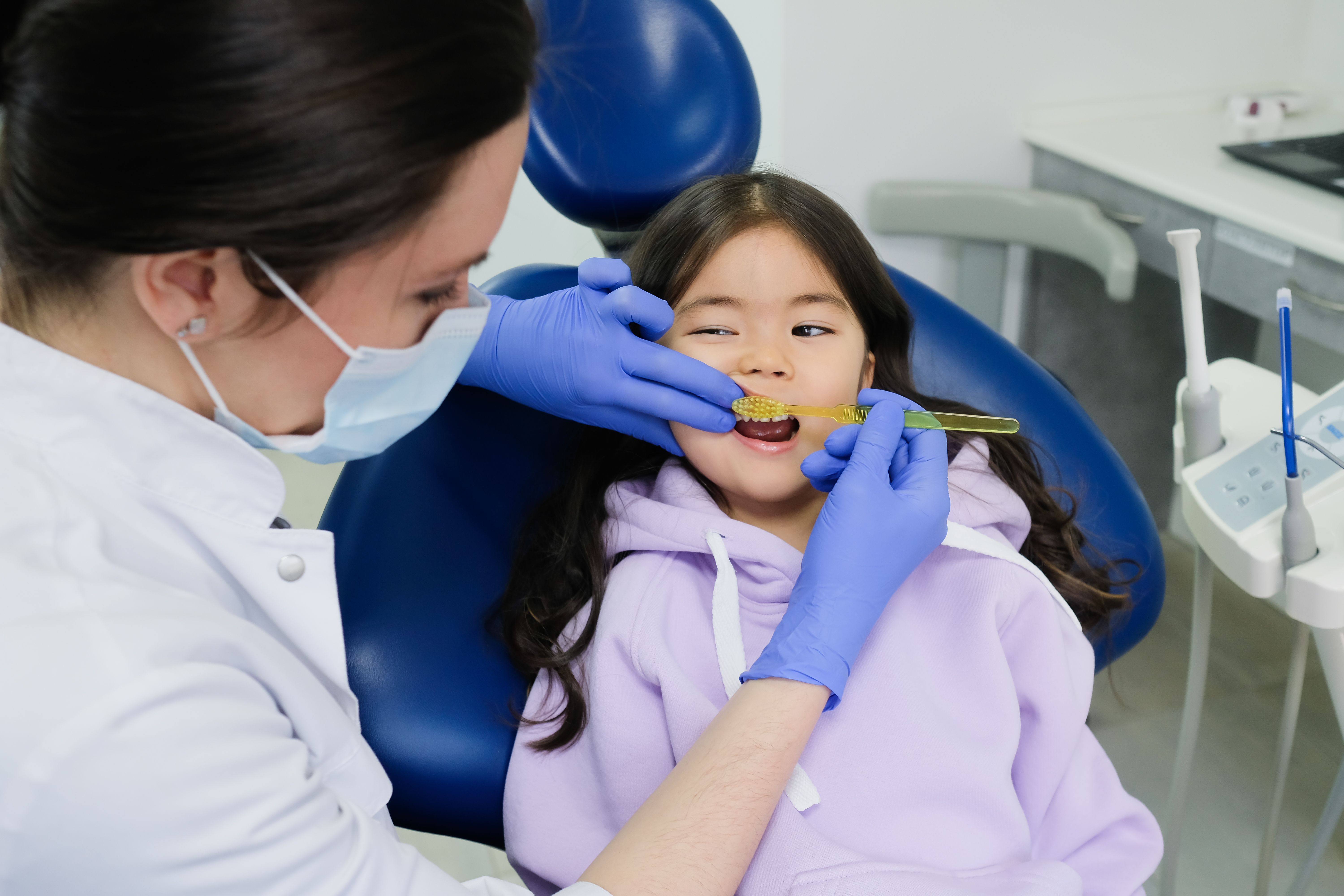
In HKDLE Part3(pediatric dental practice and licensing exams), developmental dental anomalies represent an exceptionally high-frequency testing module. In an exam setting, examiners rarely test static definitions; instead, they combine clinical photographs with radiographs to assess your diagnostic accuracy and ability to formulate comprehensive clinical treatment plans.
To help you move away from rote memorization, we have restructured these key concepts from the perspective of clinical challenges and management logic:
1. Screening for Hidden Risks: Non-Carious Periapical Lesions and Early Pulpal Exposure
Certain developmental anomalies present with subtle initial appearance but can rapidly trigger severe periapical pathology:
Occlusal Protuberance (Dens Evaginatus): Most commonly affects mandibular premolars (particularly mandibular second premolars). Its primary risk stems from the frequent presence of pulpal tissue within the tubercle, making it extremely prone to attrition or fracture. For young permanent teeth with open apices, if a fracture leads to pulpal infection, pulpal preservation or apexification/regenerative endodontic procedures should be performed according to pulp preservation guidelines.
Lingual Structural Invagination (Dens Invaginatus / Tooth within a Tooth): Most frequently seen in maxillary lateral incisors, characterized by a deep lingual pit. This invaginated channel allows bacteria to penetrate deep into the tooth structure, often leading to clinical cases where a tooth presents with a severe periapical lesion despite no visible carious defect. Diagnosis relies on radiographs or CBCT; early cases require preventive sealing, while severe cases necessitate root canal therapy or surgical intervention.
2. Differentiating Abnormally Large Crowns: Diagnostic Logic and Tooth Counting
When observing an abnormally large crown clinically or radiographically, the key lies in accurately distinguishing its developmental origin:
Fusion: Arises from the union of two originally separate tooth germs. Because it is a "two-in-one" formation, the total tooth count in the arch is usually reduced by one (assuming no supernumerary tooth is involved).
Gemination: Originates from a single tooth germ attempting to divide, resulting in a single root with two crowns. The total tooth count in the arch remains normal.
Clinical Note: If fusion occurs between a normal tooth and a supernumerary tooth, the total tooth count may appear normal. In such cases, periapical radiographs must be analyzed to evaluate root canal morphology and root structures for a definitive diagnosis.
3. Space and Occlusal Management: Delayed Eruption, Agenesis, and Microdontia
Developmental anomalies exert a profound, long-term impact on arch development and occlusal relationships, requiring a long-term management strategy:
Eruption Obstruction and Diastema (Mesiodens): Most commonly located between the two maxillary central incisors. Clinically, if delayed eruption of maxillary permanent central incisors, a large midline diastema, or retained primary teeth is observed, periapical radiographs or CBCT should be ordered immediately. If the mesiodens obstructs normal eruption or poses a cyst risk, timely surgical extraction should be planned, complemented by orthodontic traction if necessary.
Congenitally Missing Teeth (Tooth Agenesis): Excluding third molars, the most frequently missing teeth are mandibular second premolars and maxillary lateral incisors. The core focus lies in space management—such as canine substitution or future implant restoration for missing lateral incisors. If dental implants are chosen, treatment must be deferred until full skeletal maturity is reached.
Localized Microdontia (e.g., Peg-shaped Lateral Incisors): Primarily presents aesthetic and space concerns. Clinical options include minimally invasive composite resin restorations, veneers/crowns, or combined orthodontic alignment to optimize space distribution.
🎓 Exam Preparation Note: Mastering this clinical logic is crucial for passing HKDLE Part 3. Zency Education presents the Part 3 Clinical Breakthrough Course (Bilingual), led by experienced instructors. We break down real-world cases featuring 40+ high-frequency Radiographs and Clinical Pictures to help you build standardized examiner-level defense logic!
Direct message our course consultant for free study materials and trial resources!